
Lesson 1, Topic 1
In Progress
Quiz
Click Here to View Additional Images and Background
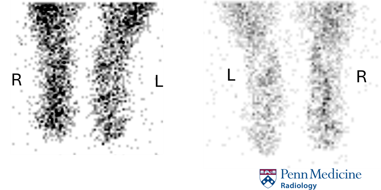
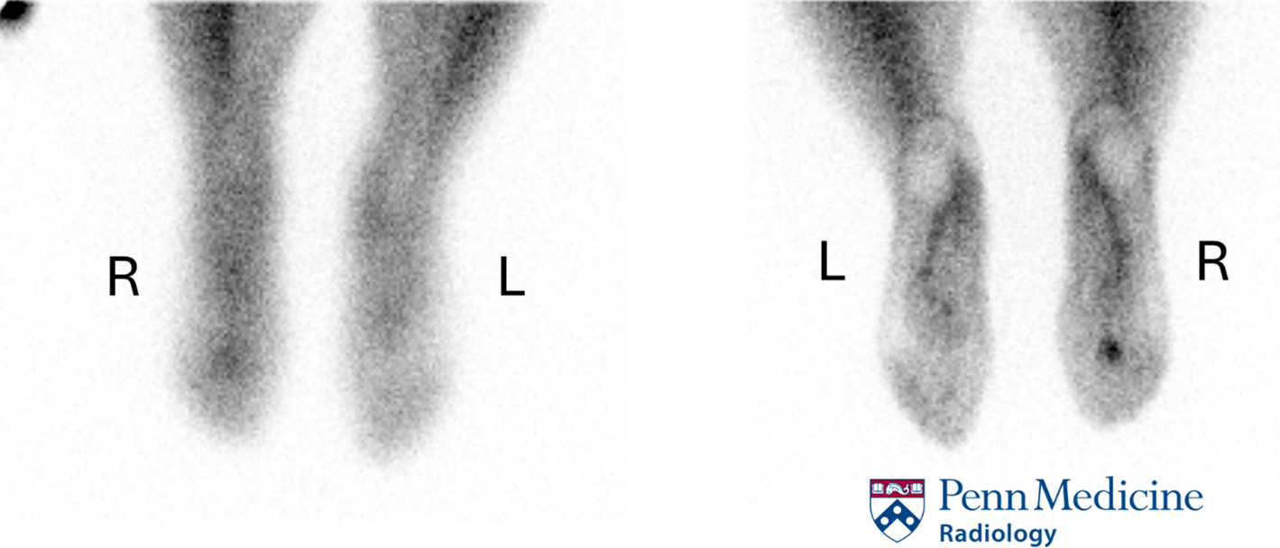
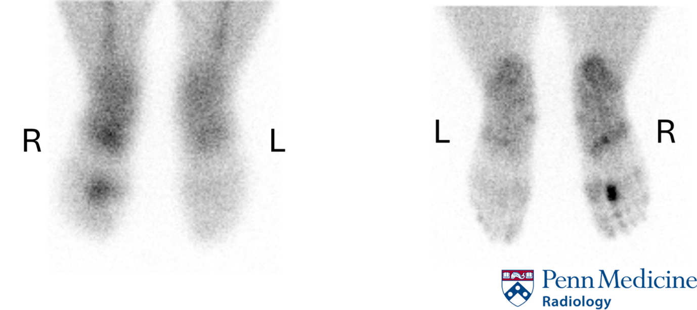
History: A 32-year-old woman presented to the outpatient physician for right foot pain. She had a history of remote hammertoe correction and was complaining of chronic pain in her right foot. She underwent a three-phase bone scan, which showed the following. Click images to enlarge.