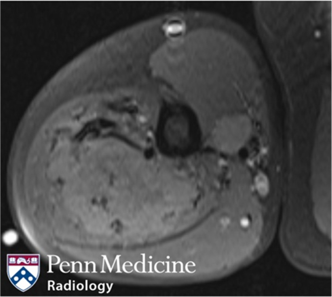
A 27-year-old woman with history of acute myelogenous leukemia (AML), status post myeloablative unrelated transplant and recent diagnosis of cutaneous…
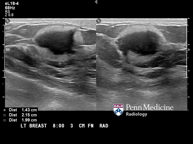
A 50-year-old woman with no significant past medical history presented with a rapidly growing tender lump in the right breast, not seen on prior scree…
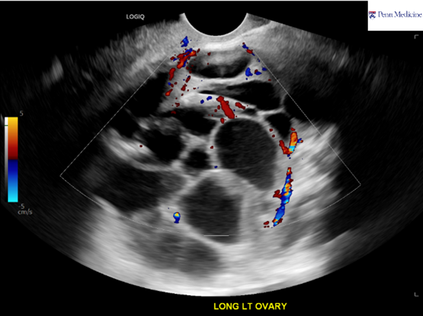
A 31-year-old woman presented to the outpatient breast imaging center for evaluation of a painless palpable lump in her right breast, persisting for t…