

Lesson 1 of 0
In Progress
Findings and diagnosis


Findings
- Unenhanced CT: A subtle hypodense lesion anterior to the intrahepatic inferior vena cava.
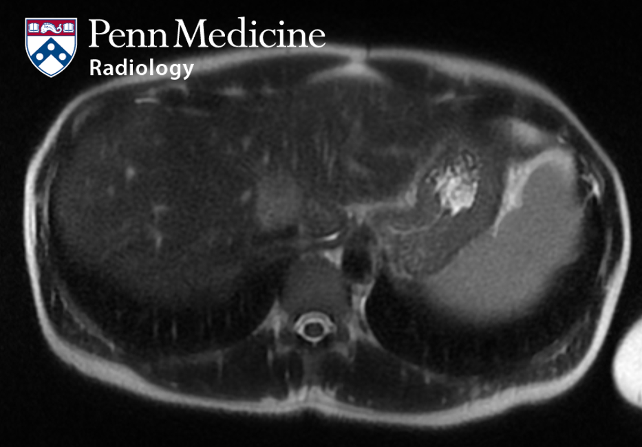
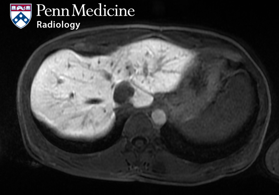
- MRI with hepatobiliary contrast: The mass measures 2.8 x 2.5 cm. This mass is T1 hypointense and T2 hyperintense relative to the liver. On dynamic postcontrast imaging, there is arterial hyperenhancement with subsequent washout on later phases. There is no enhancement on the hepatobiliary phase. There is associated mild diffusion restriction but no intracellular lipid or intralesional hemorrhage.
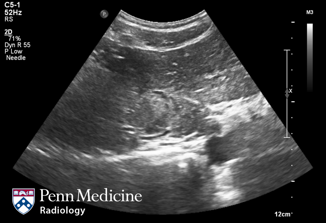
- Ultrasound: Hyperechoic, isoechoic, and hypoechoic parts relative to the surrounding liver.
Differential diagnosis
- Hepatocellular carcinoma
- Angiomyolipoma
- Hepatic adenoma
- Cholangiocarcinoma
- Metastasis
Diagnosis: Angiomyolipoma